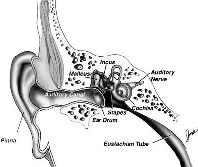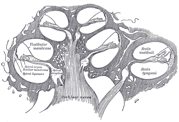
The vestibular canals of the inner ear are connected to the brain through the auditory canal, where sound waves are converted into chemical signals that are interpreted by the brain. The inner vestibular canals have a complex network of tubes and ducts that forming a canal that extends into the brain.
The inner ear consists of three small semicircular canals, each with one or more ampulla, which are air-filled sacs
The ducts lead to the brain, where they convert sound vibrations into electrical signals. The auditory canal has two primary sections: the middle and outer canals. The semicircle-shaped canals include several ampulla and some with oriented vertically and some vertically oriented horizontally.
Compared to the vertical and horizontal ampullae, the lateral ampulla is more sensitive to the relative movement of the head movements in three-dimensional (three-dimensional) space, which is possible only in comparison with the horizontal and vertical ones. The female duct system predominates in both the auditory and vestibular canals, but least of all in the funnel and ampulla.
In the ear canal, sensory nerves called the vestibulocochlear nerve lead from the sensory vestibule to the brain through the ear canal. In the vestibulocochlear vestibular reflex, there is a sudden contraction of the ear with a loud sound emitted by the brain, and the response of the auditory nerve follows the contractions of the vestibule.
The vestibule helps regulate the balance of the central auditory system. It promotes the formation of the cochlea's hearing aid. The vestibule contains a very thin and delicate bone structure called the "carpal tunnel" that separates the inner ear from the skull. It is the main barrier between the auditory bones of the cochlea and the skull.
In addition to the sensory nerves in the ear canal, there are also some muscles and cartilage in the inner ear called the vestibular muscles. which help in the movement and functioning of the vestibular sinus and vestibulocochlear duct. which allow the brain to send appropriate sound signals to and from the ears. The vestibulocochlear duct is a network of small blood vessels that are located within and around the ear canal. They promote proper drainage of the cochlea and the transmission of sound waves produced by the auditory system.

The vestibulocochlear duct does not drain directly into the middle ear but passes through a small opening in the ear called the foramen magnum before entering the inner ear. This hole is called an oval hole.
The foramen ovale collects fluid from the inner ear, which helps conduct sound waves and other nutrients to the vestibulocochlear. The fluid then moves along the cochlea and is carried out of the cochlea to the inner ear where it enters the ear drum, which contains tiny hair-like structures called vestibulocochlear endings.
These endings are responsible for stimulating the nerve cells of the vestibulocochlear, which are present in the vestibule, and they are responsible for sending impulses to the brain. When the inner ear is stimulated by a sound signal, these neurons send impulses to the brain. These impulses are interpreted by the brain, which then produces an auditory sensation.
The auditory nerves, which are located inside the ear and are connected to the vestibulocochlear by an auditory nerve, help produce the auditory sensation. As the auditory nerves and the auditory nerve to transmit their messages to the brain, the vestibulocochlear sends its signals to the brain. Thus, a hearing is affected by the stimulation of the vestibulocochlear.
The auditory nerve is a branch of the reticular motor system (RMS), which is an autonomous system in the inner ear, which transmits the information on its own to the brain to produce the sensations on the outer, central, and the internal parts of the body. The auditory nerve transmits information regarding the position and motion of the ear drum to the brain.
The afferent part of this system is not connected to the vestibule and cochlea, which are the two important structures that help generate auditory sensation. It is a motor unit that is involved in the generation of movement in the inner ear.